Join us for the next BioArt Mixer, an interdisciplinary conversation exploring how concepts of learning extend across physics, biology, narrative, and artificial systems.
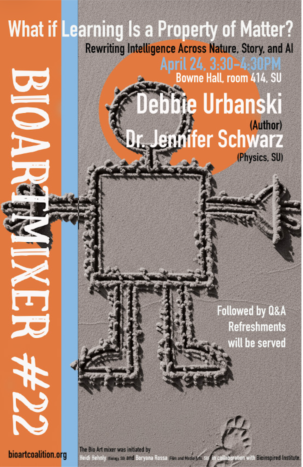
📅 April 24, 3:30–4:30 PM
📍 Bowne Hall, Room 414, Syracuse University
What if Learning Is a Property of Matter?
Rewriting Intelligence Across Nature, Story, and AI
This session features a conversation between Debbie Urbanski and Jen Schwarz, followed by Q&A and refreshments.
We often localize learning within brains—or in artificial neural networks modeled after them. This discussion instead asks whether learning may be a more fundamental property of matter itself: emerging in systems that encode history, respond to perturbation, and reorganize through interaction. Framed through both scientific and narrative lenses, the event explores how learning may be written into the fabric of physical and conceptual worlds.
Schwarz’s work in theoretical physics examines how diverse systems—from particle assemblies to cellular collectives—can encode memory and exhibit adaptive behavior through physical processes. Urbanski’s speculative writing, including After World and her recent collection Portalmania, approaches similar questions through narrative, imagining intelligence and learning beyond human-centered frameworks.
The BioArt Mixer series, initiated by Heidi Hehnly and Boryana Rossa in collaboration with the Bioinspired Institute, brings together perspectives across disciplines to foster new ways of thinking at the intersection of science and art.
All are welcome.