New Preprint: Structural Diversity and Essential Functions of Zebrafish Left-Right Organizer Cilia

by Heidi Hehnly in ,


We are excited to share our new preprint on bioRxiv, Functionally Essential and Structurally Diverse: Insights into the Zebrafish Left-Right Organizer’s Cilia via Optogenetic IFT88 Perturbation and Volume Electron Microscopy . This work was led by Favour Ononiwu (Hehnly lab Graduate student) with contributions from Melissa Mikolaj (NCI- Narayan Lab), Christopher Dell (NCI-Narayan Lab), Abdalla Wael Shamil (Hehnly lab undergraduate), Kedar Narayan (NCI), and Heidi Hehnly.

Why this study matters

During vertebrate embryogenesis, the left-right organizer (LRO) generates asymmetric fluid flow that initiates left-right body patterning. In zebrafish, this role is carried out by a transient epithelial organ known as Kupffer’s Vesicle (KV). The cilia within KV have long been known to generate flow, but their structural heterogeneity and contribution to KV morphogenesis have remained unclear.

Our approach

We combined two powerful tools:

  • Optogenetics: Using a newly engineered sox17:Cry2-GFP zebrafish line, we clustered the intraflagellar transport protein IFT88 in KV progenitors via blue-light activation. This perturbation impaired ciliogenesis and disrupted lumen formation, establishing a direct role for cilia in KV morphogenesis.

  • Volume electron microscopy (vEM): We generated the first high-resolution 3D ultrastructural map of a mature KV, enabling unprecedented analysis of ciliary architecture across the tissue.

Key findings

  • Only ~70% of cilia retained both mother and daughter centrioles, suggesting that centriole elimination may occur during KV development.

  • Among centrioles, distal appendages (34%), subdistal appendages (92%), and rootlet fibers (5%) were present in highly variable patterns, revealing remarkable structural diversity.

  • Cilia were frequently associated with membrane-bound vesicles, including ciliary-associated vesicles (CaVs) and dense vesicles (CaDVs), with distinct spatial distributions across the KV.

Broader implications

Our findings uncover previously unrecognized complexity in LRO organization. The structural specialization of KV cilia suggests that they may contribute not only to generating flow but also to organizing the architecture of the organ itself. This work adds to a growing appreciation that cilia are not uniform organelles, but instead exhibit context-specific diversity that underpins their function.

Next steps

We anticipate that these insights will inform broader studies of ciliary specialization across tissues and their role in developmental disorders linked to left-right asymmetry.

👉 Read the full preprint here.


New Publication: Dynamic Forces Sculpt Organ Shape in Zebrafish Development

by Heidi Hehnly in ,


We are excited to share our new collaborative paper with the Manning and Amack labs, published in PNAS. This work addresses a fundamental question in developmental biology: how do cells and tissues achieve the precise shapes required for organ function?

Why this matters

Many studies have focused on how cell-intrinsic properties—like signaling pathways or cytoskeletal dynamics—contribute to tissue shape. But development is more than just cells behaving individually; it is also about how tissues as a whole generate and respond to forces. Recent theoretical work suggests that embryonic tissues exist near a “jamming” transition, meaning they can flow very slowly but still transmit large forces over long timescales. These dynamic forces, though often overlooked, have been hypothesized by Manning and Amack groups to play a powerful role in shaping organs.

Our focus: Kupffer’s vesicle

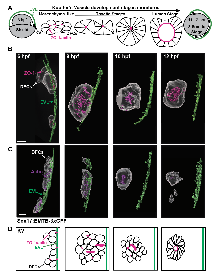
To test this idea, Amack and Manning labs turned to Kupffer’s vesicle (KV), a transient, ciliated organ in zebrafish embryos that our lab also loves to examine. KV plays a crucial role in establishing left-right asymmetry during development, making it an ideal model to study how tissues generate and respond to mechanical forces.

What we did

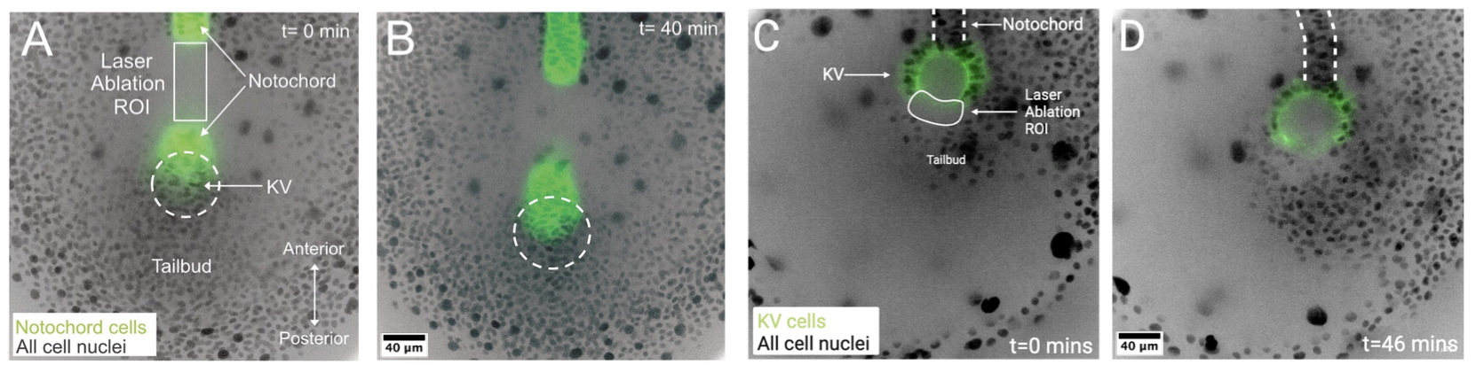
This project combined mathematical modeling, live imaging, and in vivo perturbations to test whether dynamic forces generated by tissue movements sculpt KV shape. The last part with in vivo perturbations, is where our group played an important role first with Mike Bates (a postbac in our lab and then Manager of the Blatt Imaging Center) and then with Yiling Lan (a graduate student in the lab).

  • Modeling predicted that slow tissue flows during embryogenesis could apply significant stresses to KV, driving its morphological changes.

  • Laser ablation experiments, performed in our lab (by Mike and Lan), were critical to test these predictions. By precisely severing tissue connections in the embryo, we altered force transmission and directly observed the resulting effects on KV shape. The outcomes matched the model predictions, providing strong evidence that dynamic forces are a key driver of organ morphology.

The bigger picture

The collaborative findings show that self-generated dynamic forces sculpt organ shape during development. Because many developmental processes occur on slow timescales, this principle likely applies broadly beyond zebrafish KV. This work opens the door to exploring how tissues harness dynamic mechanical forces across diverse developmental contexts.

We are thrilled to have contributed to this collaborative effort—particularly by performing the ablation experiments—and to see how interdisciplinary approaches combining modeling, physics, and cell biology can shed new light on fundamental developmental mechanisms.

📄 Read the full paper here


New Paper Alert!

by Heidi Hehnly in


Check out our new paper in Development! Titled: Specific mitotic events drive left-right organizer development.

We also did a fun interview with development titled: The people behind the papers- Yan Wu, Yiling Lan and Heidi Hehnly.

And a fun highlight of the paper can be found titled: Mitotic events help distinguish left from right.

Congrats to all the lab and everyone involved!


A new preprint from the lab!

by Heidi Hehnly in


Check out our new preprint found on bioRxiv titled: Temporal and anteriorly positioned mitotic zones drive asymmetric microtubule patterns needed for Left-Right Organizer development.

This study was a group effort led by postdoc Yan Wu and graduate students Yiling Lan and Favour Ononiwu. We were able to look at #microtubule dynamics in the #zebrafish Left Right Organizer for the first time allowing us to find lots of cool stuff about its development!

You can check it out here: https://www.biorxiv.org/content/10.1101/2024.05.12.593765v1

A teaser of how cool the MT dynamics are during zebrafish KV development.