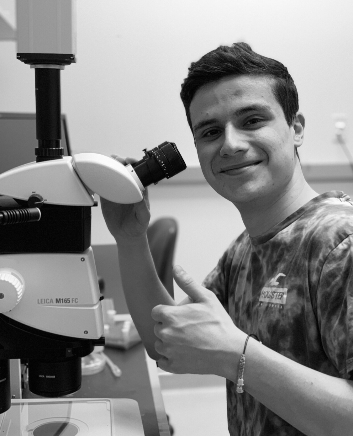
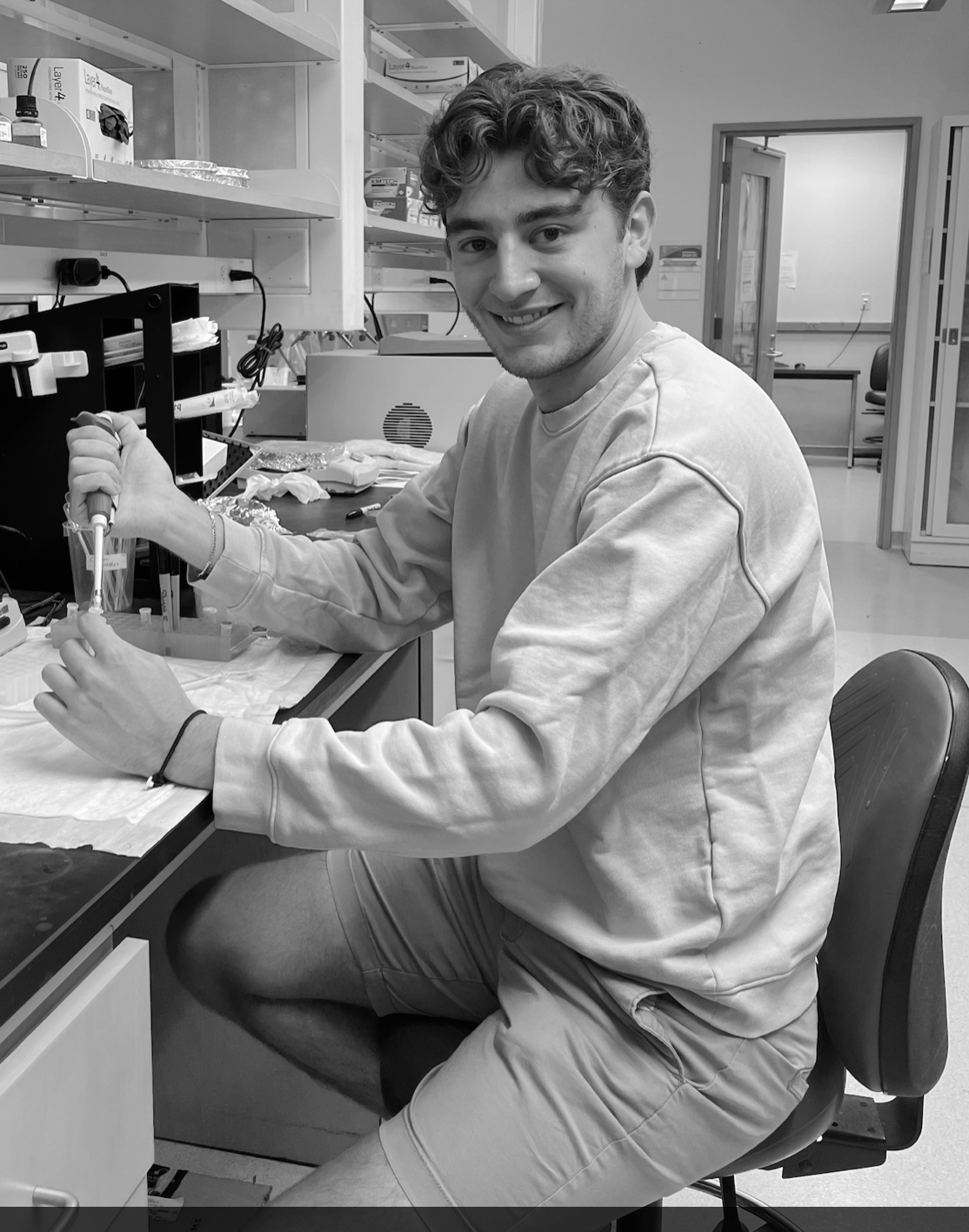
Congrats to Jonah Da Silva for being awarded the Biology Summer Research Award and SOURCE award for summer semester, and Thomas Cammerino for being awarded a SOURCE Research Award for Fall semester! Great Job!
by Heidi Hehnly
Congrats to Jonah Da Silva for being awarded the Biology Summer Research Award and SOURCE award for summer semester, and Thomas Cammerino for being awarded a SOURCE Research Award for Fall semester! Great Job!
by Heidi Hehnly in News, Microscopy Class, Lab Fun, Collaborative Work, Art
I'm so excited to help out with this Bio-Art show with Boryana Rossa and our fabulous students. If you are in the Syracuse Area, come check it out.
CHIMERA:
This exhibition features finished works and works in progress that have been made by students and artists who utilize techniques and knowledge from the field of biological sciences, apply discussion from humanities and look for visual and textual expression that comes from the arts.
During the global pandemic, the need for an integrated approach to education that includes both art and science has become imperative for fighting collective distrust in science. Our attempt is to create widely accessible view of the work done in scientific labs and open discussion about its social importance that comes from both sciences and the arts.
While taking the bio-art class the students, who have scientific or artistic backgrounds, studied examples of bio-art, had hands-on experience with microscopy and other biological techniques, and discussed their work reaching beyond their disciplines. The works in the exhibition present varieties of topics, starting with self-portraits, portraits of ecological systems, visual exploration of macro and micro worlds, ethical and personal exploration of the role of the scientist in the society, and the body as a political arena.
Guest artists are presented with signature and award-winning works. Jennifer Willet looks at the topic of co-existence, play and collaboration of human and microbial worlds, Paul Vanouse reflects upon industrial society’s shift from human and machine labor to forms of microbial manufacturing, and Adam Zaretsky presents “The Errorarium,” a device for exploring the gamification of the forced genetic errors that may appear in chamber-grown botanica.
CIMERA is part of the programming of the Bio-Art Mixer, where art and life sciences meet, faculty and grads share their research or look at it from the perspective of a different discipline. Initiated by Heidi Hehnly, Ph.D. Biology, SU; Boryana Rossa Ph.D. FMA.
Supported by CUSE Seed Grant, Department of Film and Media Arts and Department of Biology.

by Heidi Hehnly in Art, Collaborative Work, Lab Fun, Research Presentations
Come to our Bio Art Mixer Tonight in the Lundgren Room of LSC. The mixer is open to all that loves Biology and Art. What better way to spend April fools day?!

by Heidi Hehnly in Papers
Title: Rab11 endosomes and Pericentrin coordinate centrosome movement during pre-abscission in vivo
The story can be found here.
We found that conserved between the zebrafish embryo and human cells, the oldest centrosome moves in a Rab11-dependent manner towards the cytokinetic bridge sometimes followed by the youngest. Rab11-endosomes are organized in a Rab11-GTP dependent manner at the mother centriole during pre-abscission, with Rab11 endosomes at the oldest centrosome being more mobile compared with the youngest. The GTPase activity of Rab11 is necessary for the centrosome protein, Pericentrin, to be enriched at the centrosome. Reduction in Pericentrin expression or optogenetic disruption of Rab11-endosome function inhibited both centrosome movement towards the cytokinetic bridge and abscission, resulting in daughter cells prone to being binucleated and/or having supernumerary centrosomes. These studies suggest that Rab11-endosomes contribute to centrosome function during pre-abscission by regulating Pericentrin organization resulting in appropriate centrosome movement towards the cytokinetic bridge and subsequent abscission.

Differences in mitotic centrosome movement towards the cytokinetic bridge during pre-abscission between zebrafish embryos and human cells.(A) Zebrafish embryo (5 h post fertilization) with centrin-GFP (gray) and PLK1-mCh (cyan). Scale bar, 50 μm. (a’) Inset of dividing cell in (A). Time-lapse of centrin-GFP (inverted grays, top panel; grays, bottom panel) and PLK1-mCh (cyan). Video 1 Pink arrow, centrosome. Orange arrow, midbody. Dashed lines, cell boundaries. Scale bar, 10 μm. (B) Model depicting centrosome (green) movement towards the cytokinetic bridge in dividing cells within the Kupffer’s vesicle (KV) during its development. Cyan, Nucleus. Purple, Midbody. Orange, Lumen. Dark lines, KV membranes. (C) Time-lapse of a dividing cell within the KV. KV cell membranes marked with Sox17:GFP-CAAX (gray). Cyan star, rosette center. Cyan arrow, centrosome. Dashed lines, cell boundaries. Scale Bar, 10 μm. (c’) Dividing cell depicted with PLK1-mCh (fire LUT). Cyan arrow, centrosome. (D) A KV pre-abscising cell fixed and immunostained for ZO-1 (gray), γ-tubulin (cyan) and DNA (DAPI, blue). Yellow arrow, centrosome. Dashed lines, cell boundaries. Scale bar, 10 μm. (E) Number of centrosomes per pre-abscising cell with bridge directed centrosome movement calculated as both centrosomes (2 centrosomes), only one centrosome (1 centrosome) and neither centrosome (0 centrosomes) moved shown as a violin plot with median (orange) and quartiles (dark dotted lines). Two-tailed t test between Centrin-GFP and DsRed-PACT in Human (HeLa) cells (pink background). n > 10 cells across n > 3 experiments n.s. not significant. One-way ANOVA across zebrafish epiboly cells and KV cells (green background), n.s. not significant. n > 10 cells across n > 2 embryos. One-way ANOVA, across all columns, **P < 0.01. Two-tailed t test between Human (HeLa) cells and zebrafish (Epiboly, KV) cells, **P < 0.01. n-values.