Michelle, an undergraduate in our lab, gave a great lecture this past week on the pluses and minuses of widefield and laser scanning confocal microscopy in our graduate level course at SU on Microscopy Techniques in Cell Biology. She presented one of my favorite papers by Jason Swedlow that really digs into the advantages of widefield imaging with deconvolution for resolving dim fluorescent structures in live samples. The paper was titled “Measuring tubulin content in Toxoplasma gondii: A comparison of laser-scanning confocal and wide-field fluorescence microscopy” and can be found here.
Michelle presenting on Widefield Microscopy with deconvolution using the model organism Toxoplasma Gondii.
Check out Erica’s recent publication in Cytoskeleton titled “Regulating a key mitotic regulator, polo-like kinase 1 (PLK1)”. You can find the article here. Here’s her beautiful cover below, which is a Structured Illumination Microscopy Micrograph of PLK1 (Fire Look-up Table) and kinetochores (CREST, white) during different stages of the cell cycle.
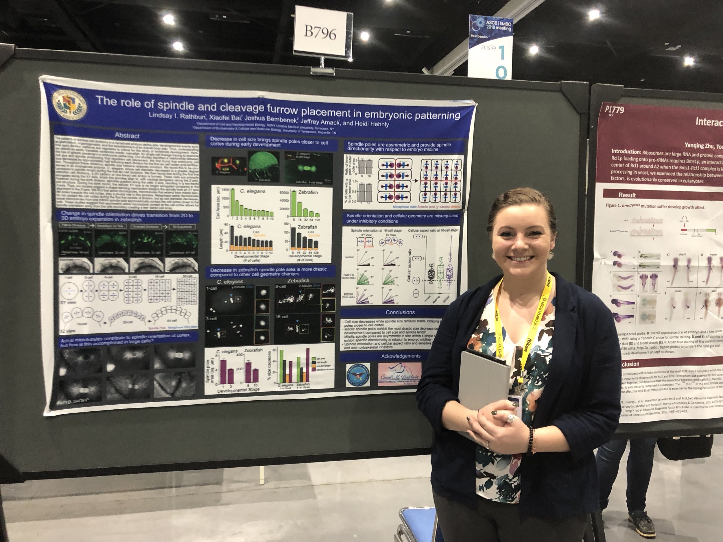
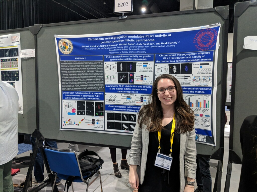
Erica Colicino, Lindsay Rathbun, and myself all presented posters this year on spindle orientation in zebrafish morphogenesis, the role of abscission in lumen formation in vivo, and chromosomes asymmetrically segregating. Also, our collaborators Carlos Castaneda and his student Julia Riley presented their work. We got lots of great feedback and got to enjoy a lot of sun. Highlights included seeing old friends from Iowa, Seattle, and Umass, and a cat cafe. Some photos below: