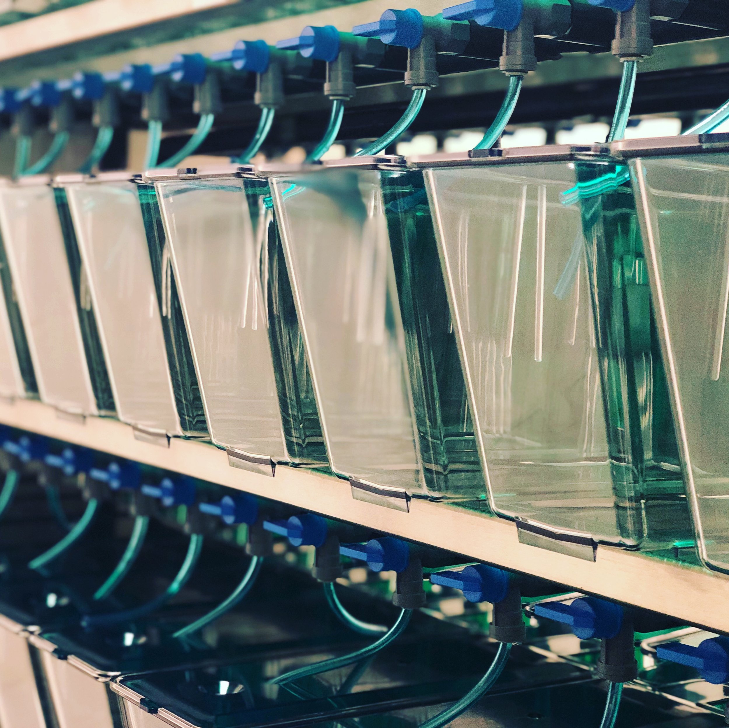
The Hehnly lab has been expanding their repertoire from 2D and 3D mammalian tissue culture to also include zebrafish as a model. We started with the help of Jeff Amack’s lab at SUNY Upstate, but now have invested in our very own zebrafish facility with the help of Aquaneering and Syracuse University. Stay tuned for two fun studies in the works where we do cell biology in the zebrafish embryo.
Graduate Student Lindsay Rathbun gave a great talk and poster at SU Biology's Graduate Student Research Day!
Lindsay at her poster in the SU life sciences complex atrium.
Lindsay at her poster in the SU life sciences complex atrium.
Michelle Nunez-Garcia giving a great lecture on the benefits between Widefield microscopy and Laser Scanning Confocal
Michelle, an undergraduate in our lab, gave a great lecture this past week on the pluses and minuses of widefield and laser scanning confocal microscopy in our graduate level course at SU on Microscopy Techniques in Cell Biology. She presented one of my favorite papers by Jason Swedlow that really digs into the advantages of widefield imaging with deconvolution for resolving dim fluorescent structures in live samples. The paper was titled “Measuring tubulin content in Toxoplasma gondii: A comparison of laser-scanning confocal and wide-field fluorescence microscopy” and can be found here.
Michelle presenting on Widefield Microscopy with deconvolution using the model organism Toxoplasma Gondii.
Congrats to Erica Colicino for her cover at Cytoskeleton
Check out Erica’s recent publication in Cytoskeleton titled “Regulating a key mitotic regulator, polo-like kinase 1 (PLK1)”. You can find the article here. Here’s her beautiful cover below, which is a Structured Illumination Microscopy Micrograph of PLK1 (Fire Look-up Table) and kinetochores (CREST, white) during different stages of the cell cycle.


