Check out the beautiful review titled “The balance between adhesion and contraction during cell division” from Dylan Burnette’s lab, specifically Nilay Taneja, that my graduate student, Lindsay Rathbun, and I were lucky to contribute to. Featured below is a stunning Figure Nilay put together. It’s fantastic and shows the power of Structured Illumination Microscopy.
Figure 1. (a) Structured illumination microscopy micrograph of HeLa cell at metaphase, stained for α-tubulin (yellow), actin filaments (magenta) and myosin IIA (cyan). The mitotic spindle comprises spindle microtubules, that facilitate chromosome segregation and dictate furrow positioning, and astral microtubules that play a pivotal role in spindle positioning by interacting with the actin cortex. Myosin II is uniformly distributed at the cortex during metaphase. (b) Upon anaphase onset, myosin II is enriched at the equator to ingress the cleavage furrow. Note the extensive contacts between the mitotic spindle and the contractile cortex, suggesting cross-talk between these two cytoskeletal networks. Note that the actin bundles protruding from the cells are not retraction fibers, as they are not attached to the substrate. Scale bar: 10 μm.
Under stress, certain eukaryotic proteins and RNA assemble to form membraneless organelles known as stress granules. The most well-studied stress granule components are RNA-binding proteins that undergo liquid-liquid phase separation (LLPS) into protein-rich droplets mediated by intrinsically disordered low-complexity domains (LCDs). Here we show that stress granules include proteasomal shuttle factor UBQLN2, an LCD-containing protein structurally and functionally distinct from RNA-binding proteins. In vitro, UBQLN2 exhibits LLPS at physiological conditions. Deletion studies correlate oligomerization with UBQLN2’s ability to phase-separate and form stress-induced cytoplasmic puncta in cells. Using nuclear magnetic resonance (NMR) spectroscopy, we mapped weak, multivalent interactions that promote UBQLN2 oligomerization and LLPS. Ubiquitin or polyubiquitin binding, obligatory for UBQLN2’s biological functions, eliminates UBQLN2 LLPS, thus serving as a switch between droplet and disperse phases. We postulate that UBQLN2 LLPS enables its recruitment to stress granules, where its interactions with ubiquitinated substrates reverse LLPS to enable shuttling of clients out of stress granules.
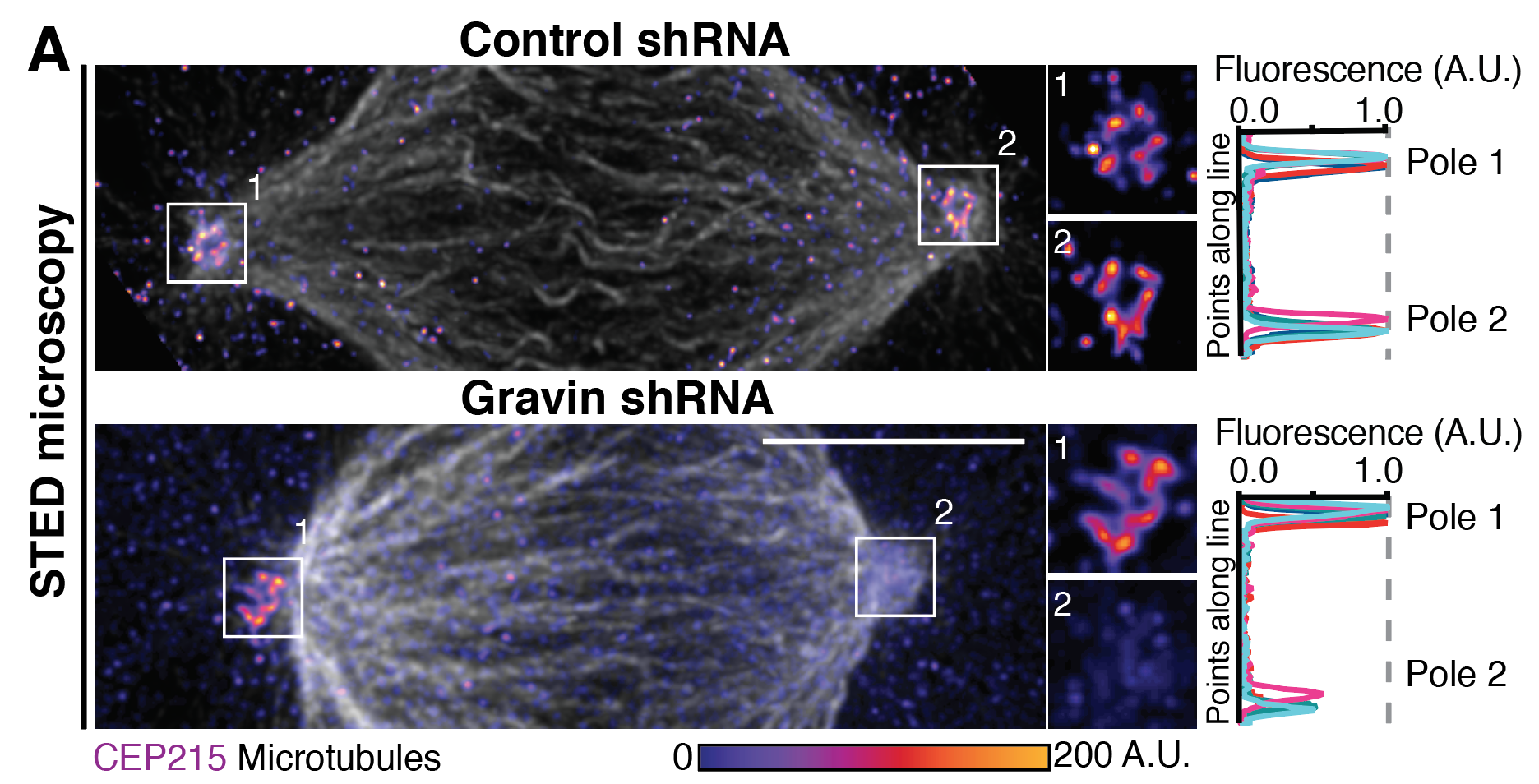
Colicino et al. talks about the role of Gravin anchoring PLK1 at mitotic centrosomes. This anchor is important for regulating centrosome organization and function as seen below with super resolution imaging of the centrosome component CEP215 disorganization when Gravin is lost.
A panel taken from Figure 4 from Colicino et al. of CEP215 disorganization primarily at a single mitotic spindle pole with Gravin loss. We argue this is through uncontrolled phosphorylation of CEP215 by PLK1 in the absence of Gravin.