Hehnly Lab Highlight: Favour Ononiwu Interview on Recent Study

by Heidi Hehnly in ,


We are excited to share a first-person interview featuring Hehnly Lab PhD graduate Favour Ononiwu, discussing her recent work on the zebrafish left–right organizer. Her study provides new structural insights into centrosomes and cilia using volume electron microscopy, advancing our understanding of how cellular architecture supports developmental patterning.

Hear directly from Favour about the scientific motivation, experimental approaches, and key findings behind this work:
👉 Read the interview


Ultrastructural Insights into Centrosomes and Cilia in the Zebrafish Left–Right Organizer

by Heidi Hehnly


We are pleased to share our recent publication, Insights into the zebrafish Left-Right Organizer's Centrosomes and Cilia via Volume Electron Microscopy, now in print in Biology Open. This study represents a collaboration between the Hehnly and Narayan laboratories and was led by recently graduated Ph.D. student Dr. Favour Ononiwu.

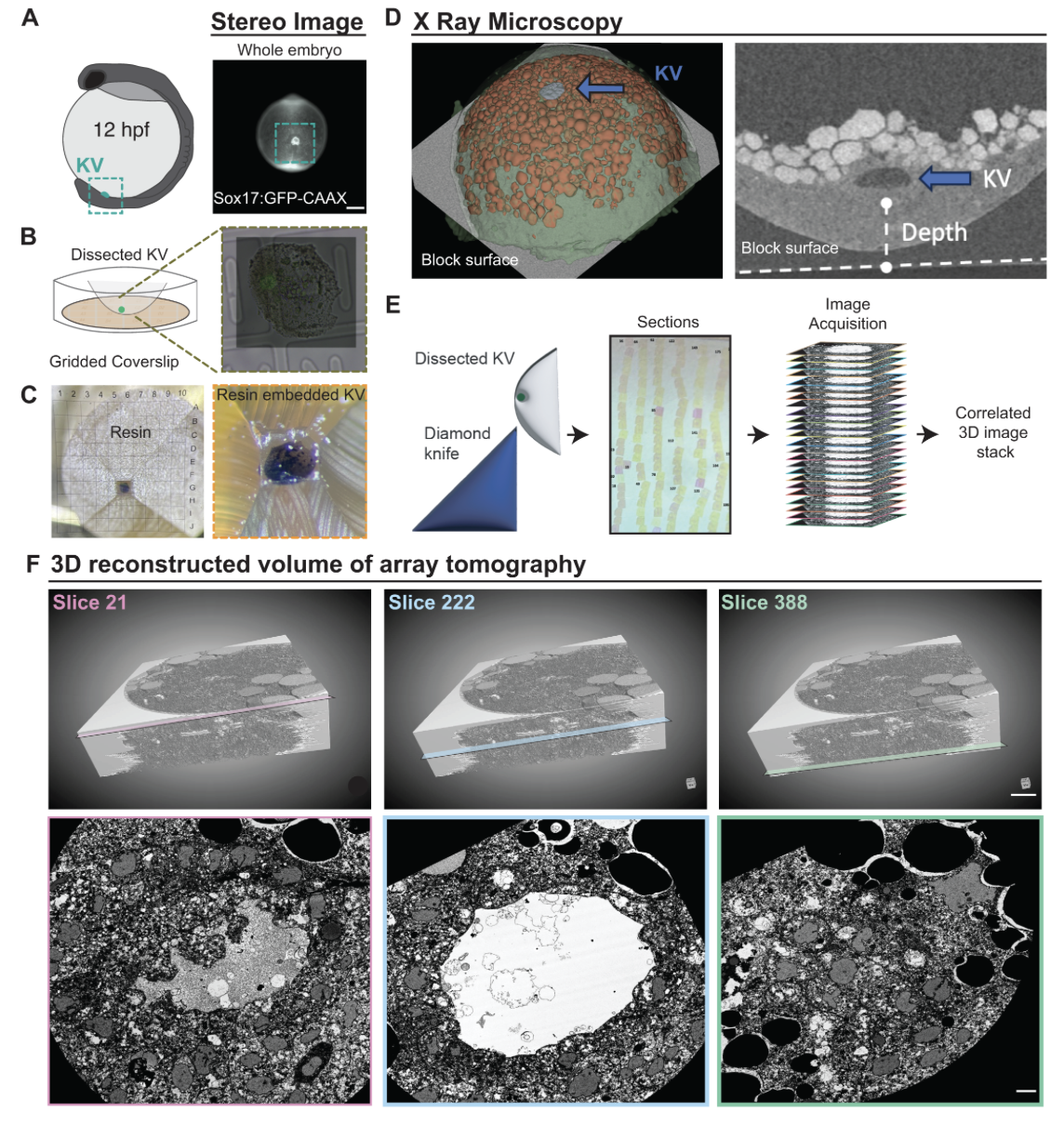
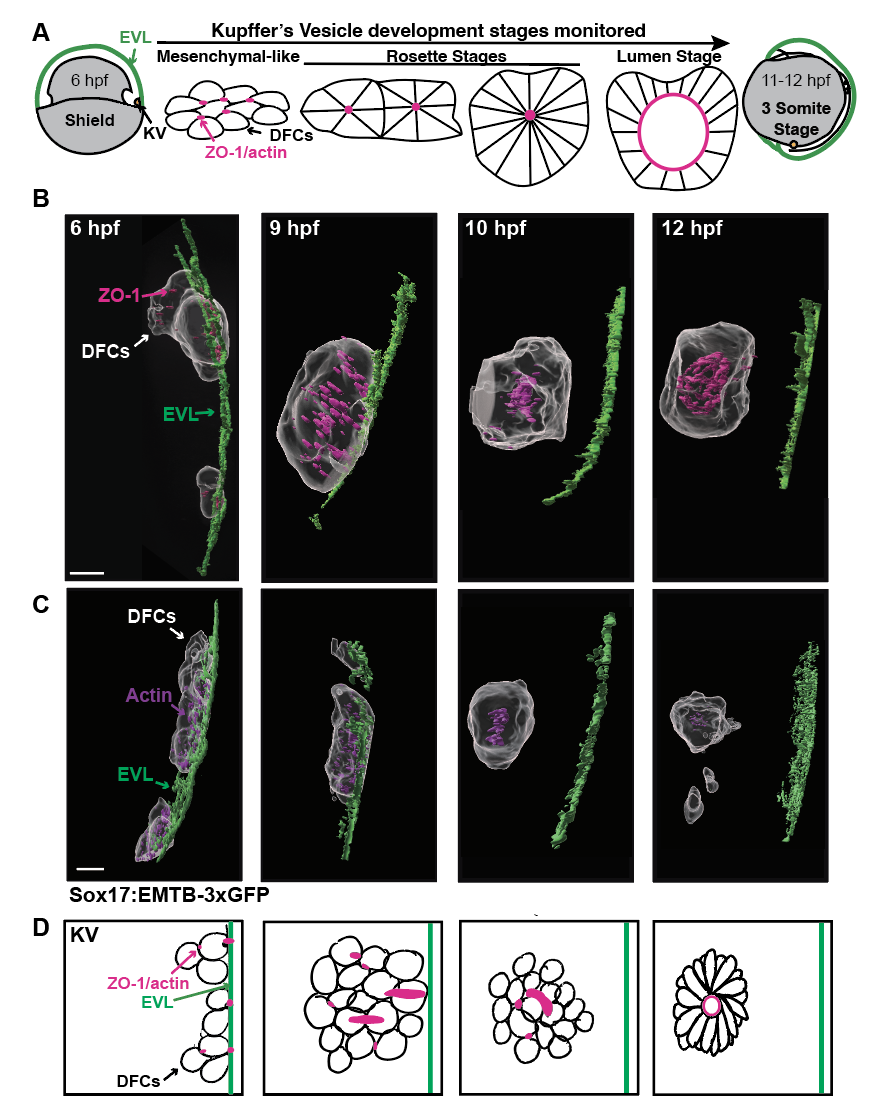
This work addresses a longstanding gap in our understanding of how ultrastructural organization within the left–right organizer contributes to symmetry breaking during vertebrate development. Using volumetric electron microscopy (vEM), we generated a near-complete three-dimensional reconstruction of the zebrafish Kupffer’s vesicle at nanometer resolution, enabling systematic analysis of cilia, centrioles, appendages, and associated vesicular structures in their native tissue context. Our findings reveal previously unappreciated heterogeneity in centrosome architecture and cilia-associated structures, including variability in centriole composition, appendage organization, and the presence of distinct vesicle populations associated with cilia.

Beyond these biological insights, this study establishes a framework for integrating volumetric ultrastructural datasets with developmental and functional analyses, providing a resource for the field and a foundation for future studies of ciliated tissues and left–right patterning mechanisms.

You can find the paper here: https://journals.biologists.com/bio/article/15/3/bio062489/371101/Insights-into-the-zebrafish-left-right-organizer-s


New Preprint: Cytoskeletal Remodeling and Junction Dynamics During KV Morphogenesis

by Heidi Hehnly in


We are pleased to share our latest preprint, now available on BioRxiv:
Read the Preprint

Understanding how epithelial architecture emerges de novo during development remains a central question in cell and developmental biology, particularly in systems where polarity and junctional organization must be established without a pre-existing template. The zebrafish left–right organizer provides a tractable in vivo context to define how transient cell–cell interactions are coordinated with cytoskeletal remodeling to drive tissue morphogenesis.

Our recent study, now available on bioRxiv, examines how dorsal forerunner cells (DFCs) dynamically remodel junctional architecture during Kupffer’s vesicle morphogenesis. Led by postdoctoral researcher Yan Wu, graduate student Yiling Lan, and exceptional undergraduate researchers Athena Allred and Carys Timpson, the work combines high-resolution imaging with quantitative analyses to show that ZO-1–based junctional assemblies form at DFC–EVL interfaces and are progressively reorganized as DFCs transition into a cohesive epithelial cyst. In parallel, we identify a role for early mitotic events and cytokinetic bridge–derived microtubule bundles in organizing actin enrichment and nucleating rosette-like structures, linking cell division to the emergence of epithelial architecture during left–right organizer development.


Welcoming New Undergraduate Researchers to the Hehnly Lab

by Heidi Hehnly in


The Hehnly Lab is pleased to welcome two new undergraduate researchers this semester, Izabella Shaina Naloy and Emily Dillon, who are contributing to ongoing projects focused on early embryonic development.

Izabella Naloy, a Biology and Forensics major, is working with postdoctoral researcher Prabhu on studies examining centriole dynamics during the earliest stages of development. Her work focuses on tracking centrioles as cells divide and reorganize during embryogenesis, an approach that will help us better understand how centrosome inheritance and positioning contribute to early developmental patterning.

Emily Dillon, an Engineering student, has joined the group working with graduate researcher Yiling Lan to investigate spindle positioning during early embryonic cell divisions. Using quantitative imaging approaches, Emily is helping measure how mitotic spindles orient and position within cells, an important determinant of cell division symmetry and tissue organization during development.

Welcome!