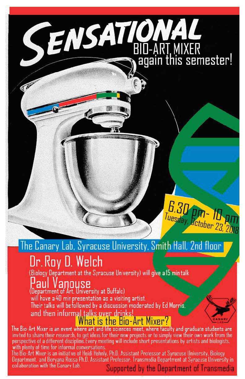
Oct 23, Tuesday, 2018
6.30-10 pm
The Canary Lab, Smith Hall, 2nd floor, Syracuse University
https://www.facebook.com/events/175580630018821/
Please make sure to spread the word in your departments and advertise! It’s great fun for students, faculty, and friends!
6.30 pm - Talks
We have two speakers Dr. Roy D. Welch (Biology Department at the Syracuse University) and Paul Vanouse (Department of Art, University at Buffalo). Dr. Roy D. Welch will give a 15 min talk and Paul Vanouse will have a 40min presentation as a visiting artist.
8.30pm- Confab
Guests’ talks will be followed by a discussion moderated by Ed Morris and by informal talks over drinks at The Canary Lab!
Paul Vanouse (Professor of Art, Director of Coalesce Center for Biological Art, Co-Director of Emerging Practices MFA, University at Buffalo)
http://www.paulvanouse.com
Paul Vanouse is an artist working in Emerging Media forms. Radical interdisciplinarity and impassioned amateurism guide his practice.
Since the early 1990s his artwork has addressed complex issues raised by varied new techno-sciences using these very techno-sciences as a medium. His artworks have included data collection devices that examine the ramifications of polling and categorization, genetic experiments that undermine scientific constructions of race and identity, and temporary organizations that playfully critique institutionalization and corporatization. These "Operational Fictions" are hybrid entities--simultaneously real things and fanciful representations--intended to resonate in the equally hyper-real context of the contemporary electronic landscape.
Roy D. Welch (Professor, Department of Biology, Syracuse University)
http://thecollege.syr.edu/people/faculty/pages/bio/Welch-Roy.html
Dr. Roy Welch studies the genetics of self-organizing behavior in the prokaryote Myxococcus xanthus through the application of molecular biology, modeling, and whole-genome analysis. Recently Dr. Welch’s lab has constructed over 100 microscopes by 3D printing to reveal how each gene in the Myxococcus xanthus genome impacts the structure and function of this communal organism to create multi-cellular structures. Dr. Welch's unique strategy in microscopic analysis of Myxococcus xanthus has been able to assign quantitative phenotype profiles to elucidate subtle functional changes linked to each gene.
What is the Bio-Art Mixer?
The Bio-Art Mixer is an event where art and life sciences meet, where faculty and graduate students are invited to share their research, to get ideas for their new projects or to simply view their own work from the perspective of a different discipline. Every meeting will include short presentations by artists and biologists, with plenty of time for informal conversations. The mixer will take place twice a semester. Our ambition is to make the Bio-Art Mixer a foundation for future exhibitions, demonstrations and new collaborative projects involving art and the life sciences, and to inspire interdisciplinary research across the universities in the region and to engage local communities.
The Bio-Art Mixer is an initiative of Heidi Hehnly, Ph.D. Assistant Professor at Syracuse University, Biology Department, and Boryana Rossa Ph.D. Assistant Professor, Transmedia Department at Syracuse University in collaboration with the Canary Lab.